Ultrasound
Ultrasound (also called sonography) is a noninvasive imaging test. An ultrasound picture is called a sonogram. Ultrasound uses high-frequency sound waves to create real-time pictures or video of internal organs or other soft tissues, such as blood vessels.
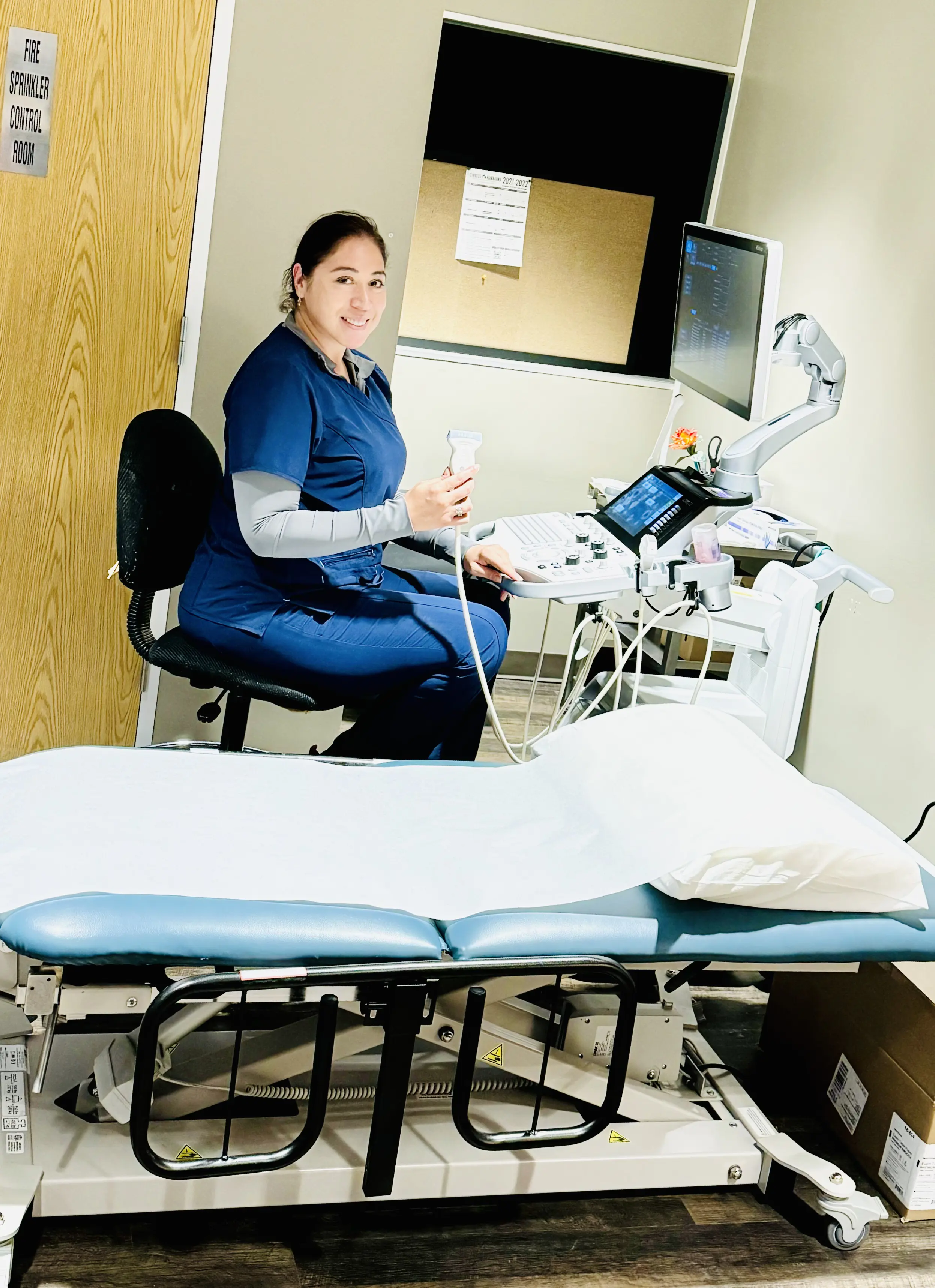
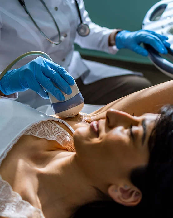
During an ultrasound, the ultrasound technician uses a device called a transducer or probe over the area of the body of interest, or inside a body opening. The tech applies a thin layer of gel to the skin so that the ultrasound waves are transmitted from the probe through the gel into the body.
The probe converts electrical current into high-frequency sound waves and sends those waves into the body’s tissue. Sound waves bounce off structures inside the body and back into the probe, which converts the waves into electrical signals. A computer then converts the patterns of electrical signals into real-time images or videos, which are displayed on a computer screen nearby.
Although most people associate ultrasound with pregnancy, it is also used to look at several different body areas such as:
- Abdominal/Stomach
- Kidneys/Renal area
- Breasts
- Pelvic
- Thyroid
- Transrectal
- Transvaginal
Campbell
9180 Katy Fwy, #100, Houston, TX 77055 | (713) 797-1919
Energy Corridor
1155 Dairy Ashford Rd, #105, Houston, TX 77079 | (713) 797-1919
Common Uses of Ultrasound
Many patients are familiar with the use of ultrasound during pregnancy. However, it also offers several other diagnostic applications. Ultrasound can detect the source of pain or inflammation inside the body and can reveal infection or tumors. It is often used for patients with suspected gallstones. Ultrasound can also evaluate the arteries and veins for narrowing, blockages, or clots.
Your Ultrasound Exam:
You will be asked to lie down on an examination table. The sonographer will apply a clear gel to the skin over the area that is being studied. The sonographer passes a small device, referred to as a transducer, over the skin. The sound waves that create the ultrasound images are sent through the transducer. You will be able to get dressed and leave immediately following the procedure. Most exams take less than 30 minutes.


