What is Nuclear Medicine?
During a nuclear medicine study, a radiotracer is typically administered through an IV, though it may occasionally be swallowed or inhaled, depending on the specific exam. This tracer travels through the bloodstream and accumulates in the specific organ or area of the body being studied, such as a tumor or an area of inflammation.
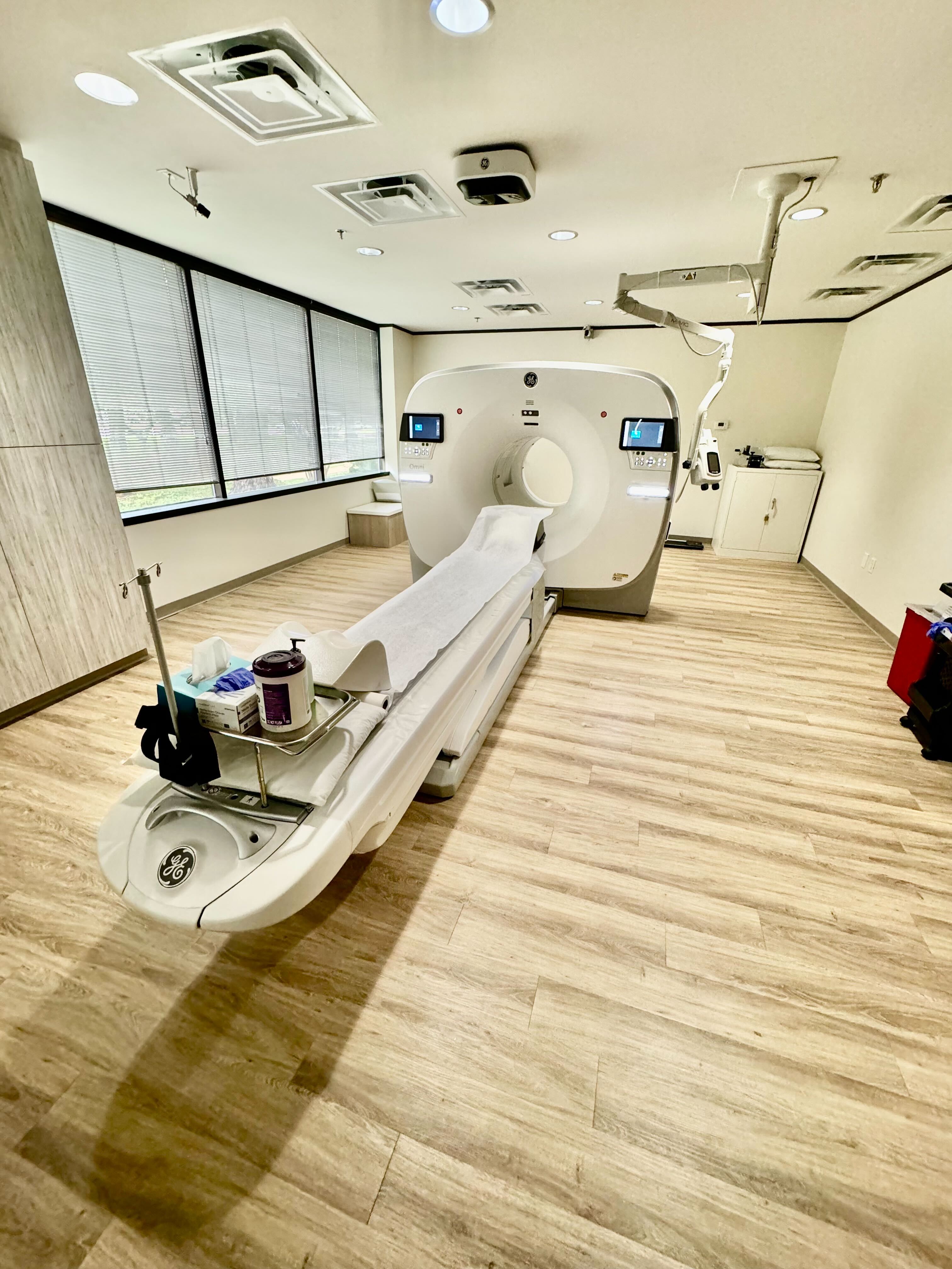
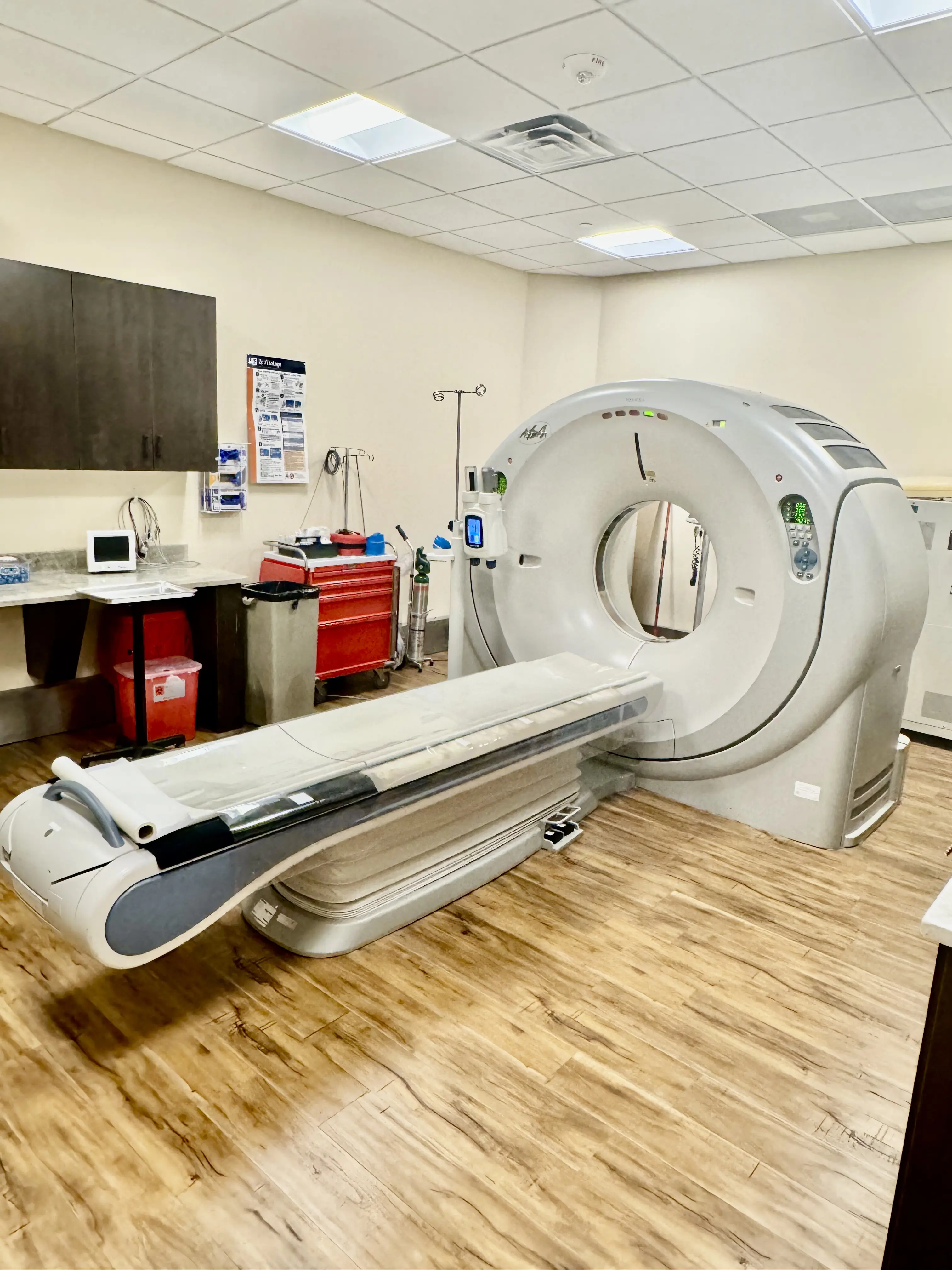
Once the tracer has reached its target, a specialized scanner, such as a PET/CT or SPECT/CT, detects the low-level radiation emitted by the tracer to create detailed computer images. While standard X-rays primarily show the structure of bones, nuclear medicine provides a "functional" view, showing our team not just what your organs look like, but how well they are working.
What Nuclear Medicine Can Detect
Nuclear medicine is a powerful tool for early diagnosis and treatment monitoring. At Houston Medical Imaging, we utilize these advanced scans to evaluate a wide range of conditions, including:
- Oncology: Detecting cancer and assessing how a tumor responds to therapy.
- Cardiology: Evaluating heart disease, blood flow, and cardiac function.
- Organ Health: Monitoring kidney, liver, and gallbladder function.
- Thyroid Disorders: Assessing thyroid activity and detecting nodules.
- Bone Issues: Identifying fractures, infections, or bone turnover that may not be visible on a standard X-ray.
Safety and Comfort
Patient safety is our top priority. Radiotracers are not dyes or medications and typically cause no side effects. The amount of radiation used is very small, often comparable to or even less than a standard CT scan. The tracer naturally decays and is flushed from your system shortly after the exam; drinking plenty of fluids following your scan can help speed up this process.