How an Arthrogram Works
An arthrogram is usually done in two steps. First, a radiologist uses live X-ray guidance (called fluoroscopy) to carefully place a small needle into the joint.
A contrast dye is then injected into the joint. This dye spreads through the area and helps highlight small tears or problems that might not be easy to see otherwise.
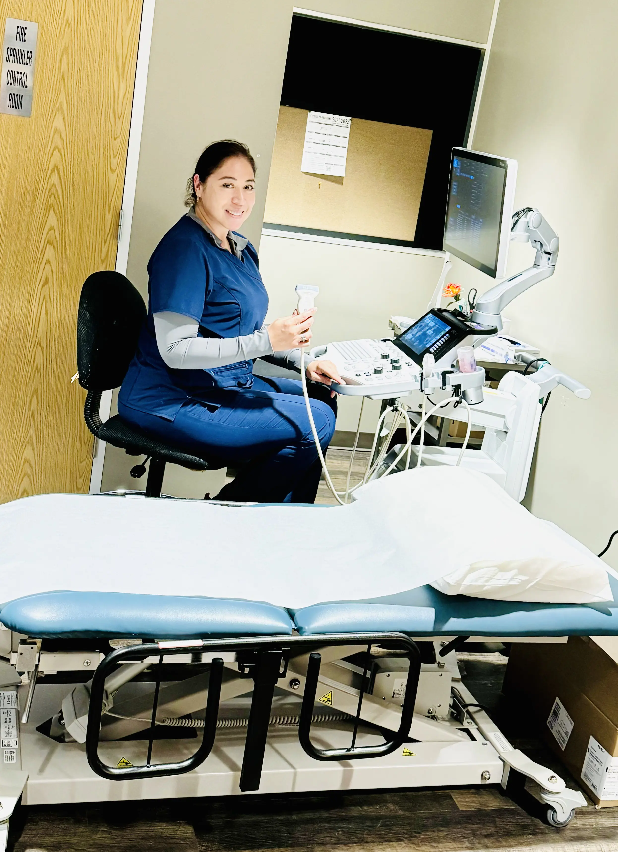
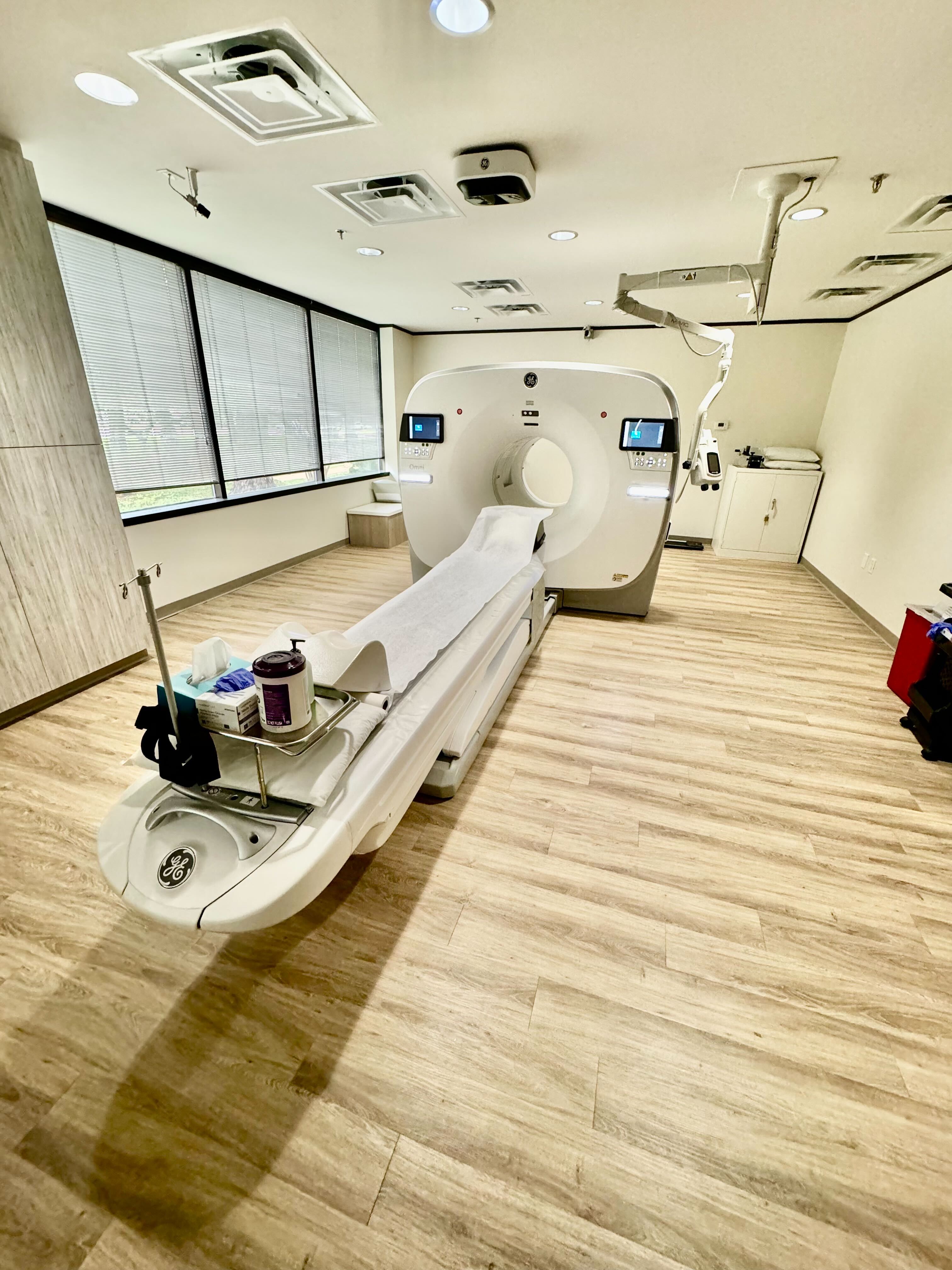
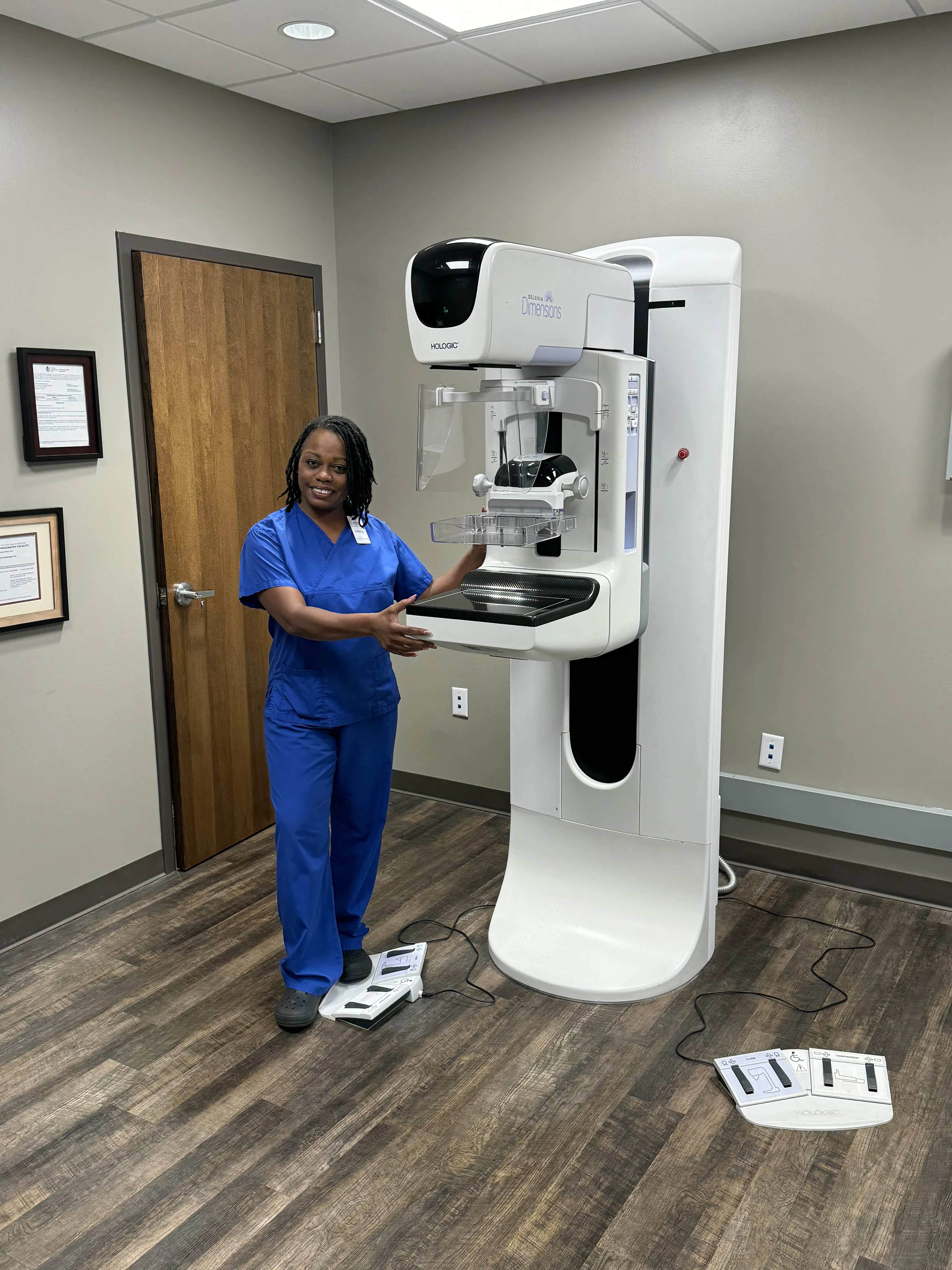
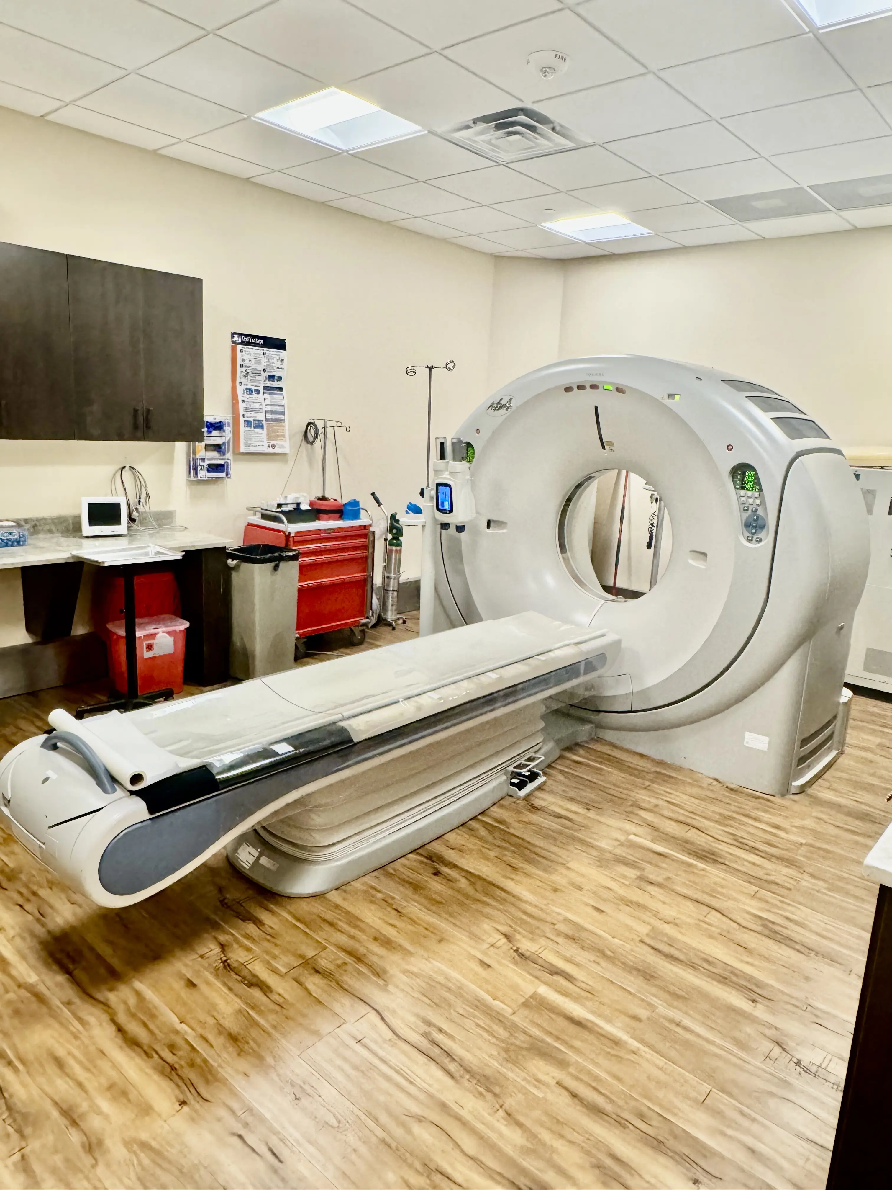
Right after the injection, images are taken using MRI, CT, or X-ray. These pictures are done quickly, while the dye is still in the joint, so your doctor gets the clearest, most accurate view.
What an Arthrogram Can Show
This procedure is most commonly used for the shoulders and hips, but it is also an important tool for checking out knees, wrists, ankles, and elbows. Our board-certified radiologists use arthrograms to:
- Diagnose Joint Pain: Find the source of unexplained pain, stiffness, or restricted movement.
- Identify Injuries: Find tears in the labrum, ligaments, cartilage, or tendons.
- Evaluate Joint Replacements: Check the condition of tissues following a joint replacement surgery.
- Therapeutic Delivery: Put a medicine, like a steroid, right into your joints to help stop swelling and pain (known as therapeutic arthrography).
What to Expect
At Houston Medical Imaging, we want you to feel as comfortable as possible during your visit. First, the area is cleaned and numbed, and a radiologist uses imaging guidance to place a small needle into the joint. You may feel a brief pinch.
A small amount of contrast dye is then injected. You might feel some pressure or fullness—this is normal. Right after, images are taken (MRI, CT, or X-ray) to get a clear view of the joint.
You can return to your normal routine afterward. Mild soreness for a day or two is common and should go away on its own.