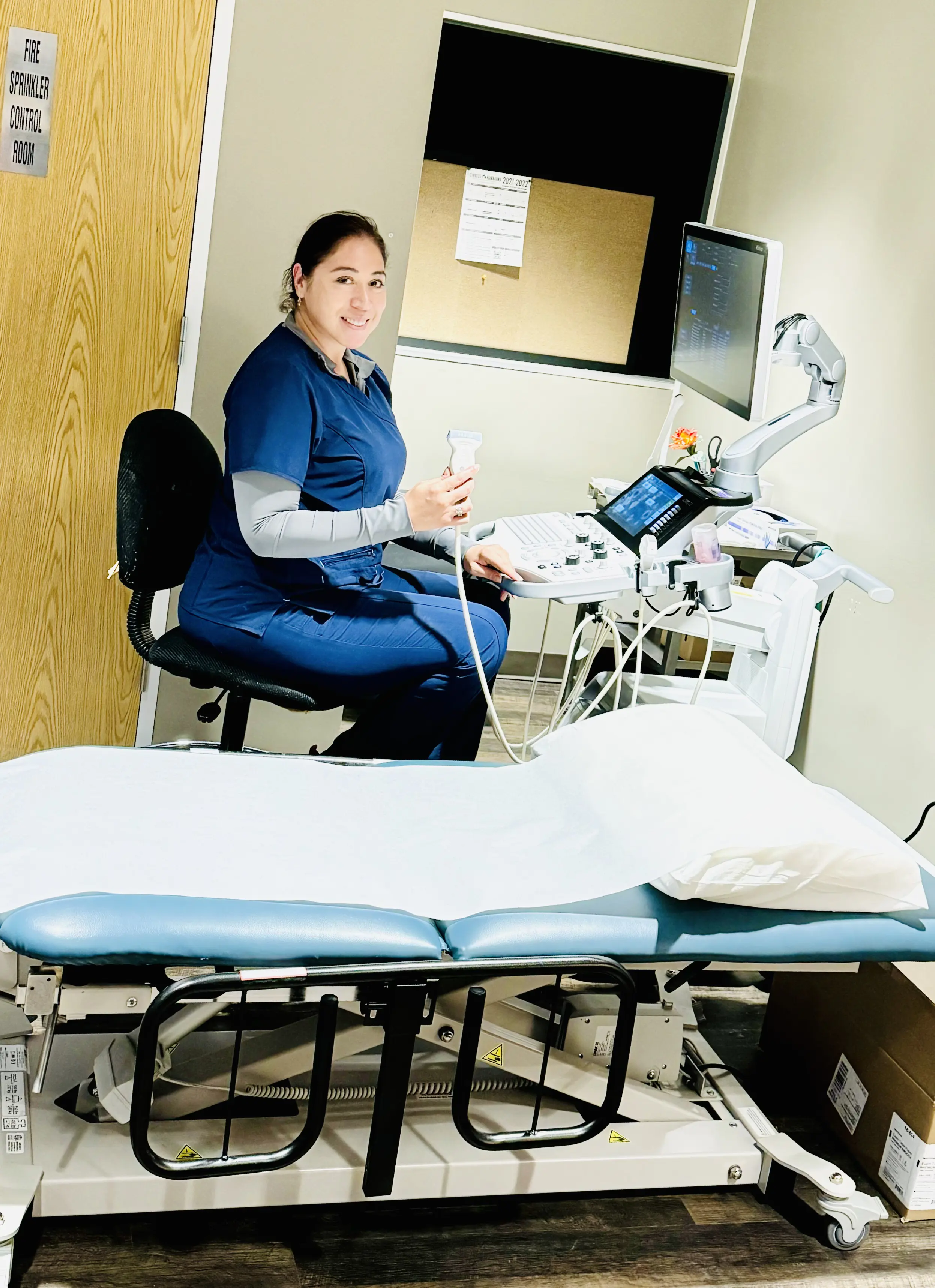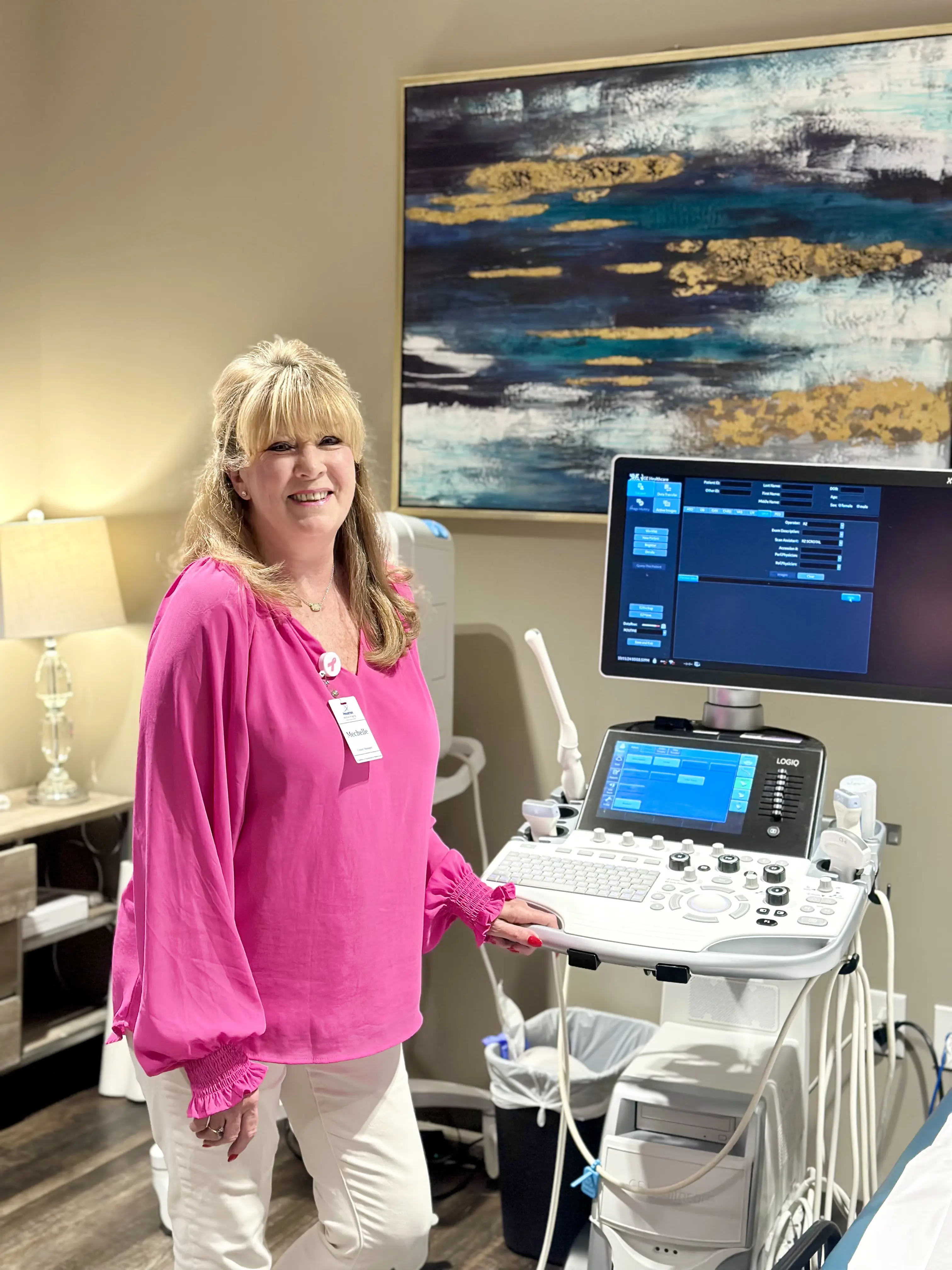
How Ultrasound Works
Ultrasound is an imaging test that uses sound waves to create pictures of the inside of your body. A small handheld device is moved over your skin, sending sound waves into the body. These waves bounce off organs and tissues, and a computer turns those echoes into real-time images. This allows doctors to see what’s happening inside your body without using radiation.
What is an Ultrasound Used For?
Most people know that an ultrasound is used to see a baby growing inside a womb. But this tool is also used for many other things. It is a very important way for doctors to check on almost any part of your body to treat medical conditions. At our 13 Houston-area locations, we use advanced ultrasound technology for:
- Abdominal Imaging: Checking the liver, gallbladder, kidneys, and bladder for stones or tumors.
- Vascular Studies: Evaluating arteries and veins for narrowing, blockages, or blood clots.
- Small Parts: Assessing the thyroid, breasts, and soft tissues for infection or inflammation.
- Pelvic Evaluations: Utilizing transabdominal or transvaginal techniques to monitor reproductive health.
Thyroid Ultrasound at Houston Medical Imaging
Our thyroid ultrasound program combines high-resolution imaging with advanced AI-supported technology to help evaluate the thyroid with greater precision and consistency.
- Detect thyroid nodules — even small or early findings
- Measure and monitor changes over time
- Evaluate features that may indicate risk
- Guide next steps, including follow-up or biopsy, if needed
What to Expect During Your Exam
Ultrasound exams at HMI Houston are designed to be efficient and stress-free. Most scans are completed in under 30 minutes. You will lie on a comfortable exam table while the sonographer gently moves the transducer over the targeted area. Once the exam is finished, you can return to your normal activities with no recovery time required.